
In vivo volumetric depth-resolved imaging of cilia metachronal wave with dynamic optical coherence tomography
Published in Optica, 2023
Recommended citation: Tian Xia, Kohei Umezu, Deirdre M. Scully, Shang Wang, and Irina V. Larina, "In vivo volumetric depth-resolved imaging of cilia metachronal waves using dynamic optical coherence tomography," Optica 10, 1439-1451 (2023) https://opg.optica.org/optica/abstract.cfm?doi=10.1364/OPTICA.499927
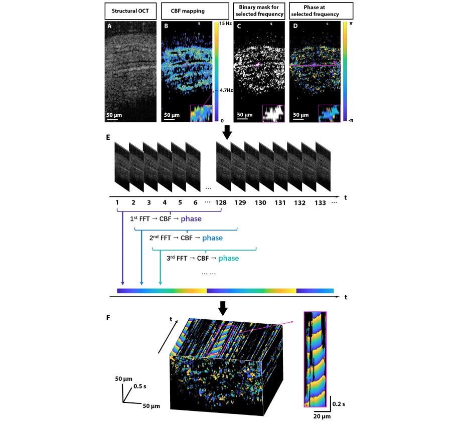
Motile cilia are dynamic hair-like structures that cover epithelial surfaces in multiple organs. The periodic coordinated beating of cilia creates waves propagating along the surface, called the metachronal waves, which transport fluids and mucus along the epithelium. Motile ciliopathies arise due to disrupted coordinated cilia beating and are associated with serious clinical complications including reproductive disorders. Despite the recognized clinical significance, investigations of cilia dynamics are extremely limited. Here, we present the first quantitative imaging of cilia metachronal wave volumetrically through tissue layers with dynamic optical coherence tomography. Our method relies on spatiotemporal mapping of the phase of intensity fluctuations in OCT images due to the ciliary beating. We validated our new method ex vivo and implemented it in vivo to visualize cilia metachronal wave propagation within the mouse fallopian tube. This new method can be extended to the assessment of physiological cilia function and ciliary dyskinesias in various organ systems, contributing to better management of pathologies associated with motile ciliopathies.
Download Accepted Manuscript here
Recommended citation: Tian Xia, Kohei Umezu, Deirdre M. Scully, Shang Wang, and Irina V. Larina, “In vivo volumetric depth-resolved imaging of cilia metachronal waves using dynamic optical coherence tomography,” Optica 10, 1439-1451 (2023).